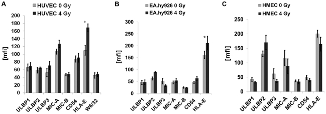
Figure 6. Phenotyping of ECs before and after irradiation at 4 Gy.
Comparative analysis of the mean fluorescence intensity values for ULBP1-3, MIC-A/-B, CD54 (ICAM-1) and HLA-E expression on HUVECs (A), EA.hy926 cells (B) and HMECs (C) after ionizing irradiation at the sub-lethal dose of 4 Gy followed by a recovery period of 12 h. The MHC class I expression, as determined by W6/32 mAb, was measured only on HUVECs. The expression density of HLA-E was found to be significantly reduced on HUVECs and EA.hy926 cells (*, p<0.05), but not on HMECs following irradiation. No significant changes were observed with respect to other cell surface markers such as ULBP1-3, MIC-A/-B, CD54 and MHC class I antigens.

