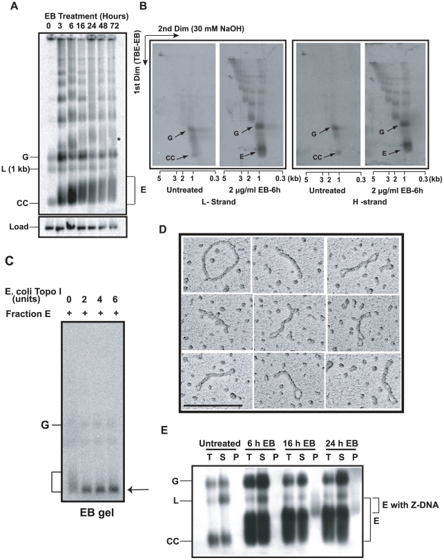
Figure 2. Discovery and characterization of Fraction E.
(A) Effect of EB on free minicircle population. Total DNA (5×10⁶ cell equivalents/lane), from cells treated with 2 µg/ml EB for indicated time, was fractionated on an agarose-EB gel. The autoradiograph is a Southern blot probed for minicircles and hexose transporter gene (load). G, gapped, L, linearized, and CC, covalently-closed minicircles. E is Fraction E. * is probably interlocked minicircle dimer forming a smear like that of Fraction E. We usually see only trace amounts of linearized minicircles in untreated cells (0 time point). (B) Two-dimensional neutral-alkaline gel electrophoresis of free minicircles. The sample was total DNA (5×10⁶ cell equivalents/lane), from untreated or EB-treated (6 h, 2 µg/ml) cells. 32P-labeled oligonucleotides were used for probing minicircle L (left two panels) and H (right two panels) strands. Scales below gels are sizes of linear markers. Abbreviations of minicircle species are same as in Panel A. See [25], [26] for description of other bands in these gels. (C) Treatment with Topo I. Fraction E (10 µl, sucrose gradient purified) was treated with E. coli topo I [51] and fractionated on an agarose-EB gel. Arrow indicates position of covalently-closed relaxed minicircles. (D) EM of fraction E also sucrose gradient purified. Molecule at upper left is a relaxed minicircle. Scale bar, 200 nm. (E) Fraction E contains regions of Z-DNA. Total DNA (T) from 50 ml of culture (0.8×10⁶ cells/ml) of untreated or EB (2 µg/ml)-treated cells was immunoprecipitated with anti-Z DNA antibody and then with protein G-Sepharose [24]. Upon centrifugation, DNA in supernatant (S) and pellet (P) was electrophoresed and probed for minicircles. Increases in linearized minicircles could be due to nuclease contamination of the antibody. Gel conditions and abbreviations of minicircle species are same as in Fig. 2A.

