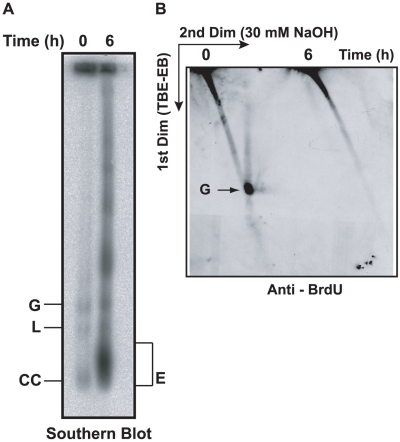
Figure 3. Effect of EB treatment on replication of minicircles.
(A) Cells (50 ml, 0.7×10⁶ cells/ml, treated with 2 µg/ml EB for 6 h) were incubated with 50 µM BrdU during the last 40 min. Free minicircles were then fractionated on an agarose-EB gel as in Fig. 2A, and a Southern blot was probed for minicircles. (B) The same amount of DNA used for the gel in Panel A was run on a 2D neutral/alkaline gel (as in Fig. 2B) and a blot was probed with anti-BrdU antibody. BrdU label at top of gel is in nuclear DNA. Abbreviations of minicircle species are same as in Fig. 2A.

