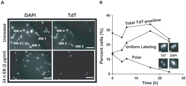
Figure 6. Effect of EB on distribution of gapped circles in isolated networks.
Isolated networks were labeled by fluorescein-12-dUTP using terminal deoxynucleotidyl transferase (TdT) [33]. This procedure adds a fluorescent tag to 3′ OH groups flanking minicircle gaps. Because of the relative abundance of minicircles and the distribution of fluorescein fluorescence within networks, most gaps must be in minicircles rather than maxicircles [33]. This procedure reveals the extent of replication of a network. TdT-positive networks are mostly undergoing replication and some are post-replication with gaps yet to be repaired. In wild type cells, the labeling pattern is usually polar or uniform, representing early and late stages of replication respectively. (A) Fluorescent images of networks isolated from untreated cells stained with 2 µg/ml DAPI (upper-left panel) and labeled with TdT (upper-right). Lower panels are the same except cells were EB-treated (2 µg/ml, 24 h). In upper panel, network (NW) 1 is a TdT-negative pre-replication or post-replication network because it stains with DAPI but not TdT. Networks 2 and 3 are replicating networks with polar labeling. Network 4 is TdT-positive with uniform labeling, appearing double-size and ready to divide. Scale bars, 5 µm. (B) Kinetics of change in TdT labeling pattern during EB treatment. Upper inset shows a uniformly labeled network and lower inset shows one with polar labeling. At least 120 networks were counted at each time point. Scale bars, 1 µm.

