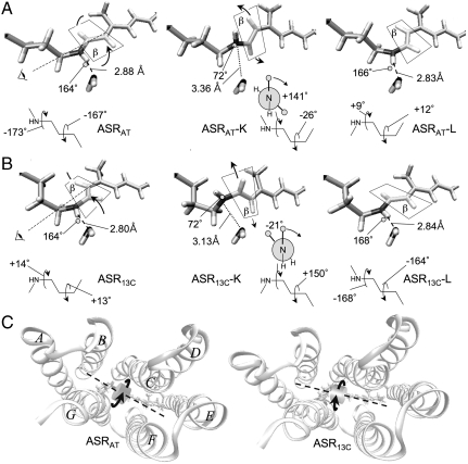
Fig. 3.
(A and B) Structural evolution of the retinal chromophore during the photochromic cycle of ASR. The crystallographic water molecule (W402) in the vicinity of the Schiff base nitrogen is also shown, together with the corresponding N─H---O distances and N─H---O angles. The ASRAT → ASRAT-K and ASR13C → ASR13C-K transitions occur with the same mechanism corresponding to a ca. 180° rotation of the plane containing the ═C14─C15═ fragment (plane β). As indicated by the dashed rotation axis and “eye” symbol in the ASRAT and ASR13C structures, this rotation occurs CCW with respect to the Lys210 side chain. The N-H bond undergoes a large and reversible reorientation due to the change in ─C15═N─Cε─Cδ─ dihedral angle that accompanies the isomerization of the ─C13═C14─ bond. During the subsequent K → L transition, the N-H bond rotates in the reverse direction, bringing about the isomerization of the ─C15═N─ bond. This backward rotation is driven by the reconstitution of the N─H---O hydrogen bond. The Newman projections display the pretwisting of the ─C15═N─ bond, which clearly favor CW isomerization of this bond during the K → L transition. The evolution of the ═C14─C15═NH─Cε─ and ═C12─C13═C14─C15═ dihedrals describing the two isomerizations is reported below each structure in degrees. (C) Top (i.e., cytoplasm side) view of the orientation of the “rotating” ═C14─C15═ moiety in ASRAT and ASR13C. The retinal backbone axis is represented by a dashed line.

