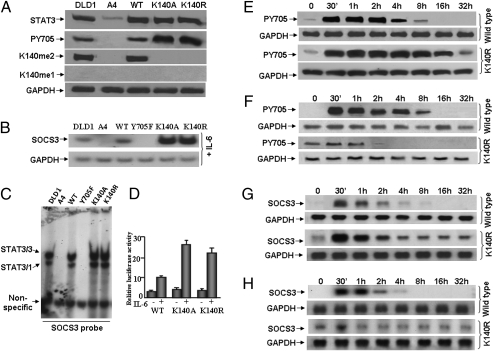
Fig. 1.
K140A or K140R mutation of STAT3 increases its tyrosine phosphorylation and transcriptional activity in response to IL-6. (A) Western analyses for STAT3 and Y705-phosphoryl-STAT3. STAT3-null A4 cells were infected with retroviral constructs and stable pools of cells were selected with G418. DLD1, parental colon cancer cells; A4, STAT3-null cells; WT, A4 cells expressing a normal level of wild-type STAT3; K140A, A4 cells expressing a normal level of K140A STAT3; K140R, A4 cells expressing a normal level of K140R STAT3. The cells were treated with IL-6 for 4 h and total cell lysates were analyzed by Western blot. (B) Northern analysis of gene expression. Cells were treated with IL-6 for 4 h and total RNA was analyzed for GAPDH and SOCS3 mRNAs. (C) EMSAs. Whole-cell extracts were made from DLD1 cells, A4 cells, or A4 cells expressing wild-type, Y705F, K140A, or K140R STAT3, treated with IL-6 for 4 h. A GAS (STAT3 binding) element derived from the SOCS3 promoter was used as the probe. (D) SOCS3 reporter gene induction. Luciferase constructs containing ∼1 kb of the human SOCS3 promoter (from −1 to about −1,000) were cotransfected with a pCH110 control plasmid. Cells stimulated with IL-6 for 4 h or untreated cells were washed with serum-free medium and cultured in complete medium for 12 h more. Cell lysates were analyzed for luciferase activity, which was normalized to the level of β-galactosidase activity from pCH110 control cells in the same extract. Values are means of triplicate determinations and the bars show one SEM. (E) A4 cells expressing wild-type or K140R STAT3 were treated with IL-6 for 4 h. The cells were washed with serum-free medium twice and cultured in complete medium without IL-6. Whole-cell lysates were analyzed by Western blot for total STAT3 and Y705-phosphoryl-STAT3. (F) A4 cells expressing wild-type or K140R STAT3 were treated with IL-6 for 4 h. The cells were washed with serum-free medium twice and cultured in complete medium without IL-6. After 32 h, the above cells were retreated with IL-6 for 4 h, and then washed with serum-free medium twice and cultured in complete medium, again without IL-6. Whole-cell lysates were analyzed by Western blot for total STAT3 and Y705-phosphoryl-STAT3. (G) A4 cells expressing wild-type or K140R-STAT3 were treated with IL-6 for 4 h. The cells were washed with serum-free medium twice and cultured in complete medium without IL-6. Total RNA was analyzed for GAPDH and SOCS3 mRNAs. (H) A4 cells expressing wild-type or K140R STAT3 were treated with IL-6 for 4 h. The cells were washed with serum-free medium twice and cultured in complete medium without IL-6. After 32 h, the cells were retreated with IL-6 for 4 h, washed with serum-free medium twice and cultured in complete medium without IL-6. Total RNA was analyzed for GAPDH and SOCS3 mRNAs.

