Fig. 8.
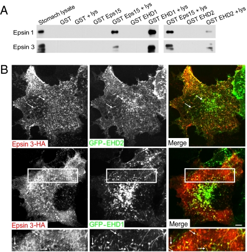
EHD1 and EHD2 interact with epsin. (A) Anti-epsin 1 and 3 Western blots of material affinity purified by GST or GST fusions of the EH domains of Eps15 and of EHD1 or EHD2. The starting lysate and the pellets obtained by incubating each fusion protein with and without lysate (lys) are shown. (B) Fluorescence images of COS-7 cells cotransfected with HA-tagged epsin 3 and GFP-EHD proteins. The lower panels shows the boxed area of middle panels at higher magnification. Arrows indicate some structures positive for both epsin 3 and EHD proteins. (Scale bars, 10 μm.)

