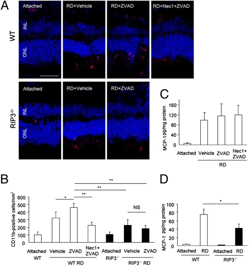
Fig. 6.
Effect of RIP pathways on inflammatory response after retinal detachment. (A and B) Immunofluorescence for CD11b (A) and quantification of CD11b-positive macrophage/microglia in WT and Rip3−/− retina after retinal detachment. In WT mice, Z-VAD treatment significantly increased infiltration of CD11b-positive cells compared with vehicle treatment (P < 0.05). This increase of CD11b-positive cells was significantly suppressed with Nec-1 plus Z-VAD treatment or Rip3 deficiency (P < 0.01). (Scale bar, 50 μm.) (C and D) ELISA for MCP-1 on day 3 after retinal detachment in retina treated with vehicle (n = 5), Z-VAD (n = 5), or Nec-1 plus Z-VAD (n = 6) (C) and in WT and Rip3−/− retina (n = 5 each) (D). The retinas without retinal detachment were used as controls. *P < 0.05; **P < 0.01; NS, not significant.

