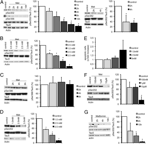
Fig. 2.
Metformin induces dephosphorylation of tau at PP2A-sensitive sites. Representative Western blots and quantifications of three or four independent experiments are shown in A–D, F, and G. Band intensities were normalized to actin and compared with total tau (Tau 5). (A) Primary cortical neurons of wild-type mice were treated with 2.5 mM metformin (Met) over increasing time intervals. Cell lysates were analyzed by Western blot using antibodies detecting phosphorylation at specific PP2A-sensitive tau sites (Ser202, Ser262, Ser356; n = 3). (B) Primary cortical neurons from wild-type mice were treated with increasing concentration of metformin over 4 h. Cell lysates were analyzed by Western blot using an antibody detecting phosphorylation at Ser202 (n = 4). (C) Primary cortical neurons of wild-type mice were treated with 2.5 mM metformin over increasing time intervals. Cell lysates were analyzed by Western blot using an antibody detecting phosphorylation at the PP2A-insensitive tau site Ser396 (n = 3). (D) Primary cortical neurons of transgenic mice expressing human tau instead of murine tau were treated with increasing concentration of metformin over 4 h. Cell lysates were analyzed by Western blot using an antibody detecting phosphorylation at Ser202 (n = 3). (E) TUNEL assay of primary cortical neurons of wild-type mice treated with increasing concentrations of metformin. Percentages of apoptotic cells are shown. (F) Primary cortical neurons of wild-type mice were treated with low concentrations of metformin over 24 h. Cell lysates were analyzed by Western blot using an antibody detecting phosphorylation at Ser202 (n = 3). (G) Primary cortical neurons of wild-type mice were treated with 2.5 mM metformin over increasing time intervals in medium without insulin. Cell lysates were analyzed by Western blot using an antibody detecting phosphorylation at Ser202 (n = 3). *P < 0.05.

