Fig. 2.
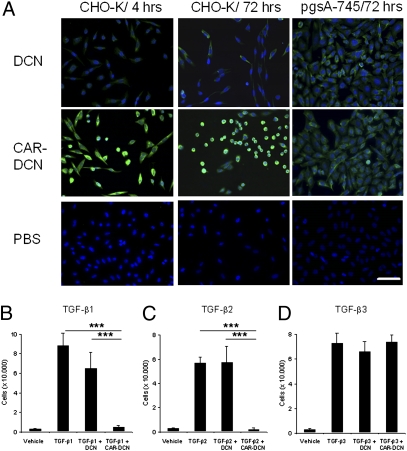
Cell binding and inhibition of cell spreading and TGF-β–dependent cell proliferation by decorins. (A) The culture media of CHO-K and pgsA-745 cells were supplemented with 0.3 μg/mL of decorins daily for 4 h or 3 d. After washing, the decorins were detected with anti–his-tag antibody and FITC-conjugated secondary antibody (green). The CAR-targeted decorin bound to CHO-K cells more (4 h) and inhibited cell spreading more strongly (3 d) than unmodified decorin, whereas the two decorins show identical low binding to mutant pgsA-745 cells that lack the receptor for the CAR peptide (19). Cell nuclei were stained by DAPI (blue). (Scale bar, 50 μm.) (B) CHO-K cells were grown in the presence of TGF-β1, (C) TGF-β2, or (D) TGF-β3 (all at 7.5 ng/mL) and 0.3 μg/mL of decorins. Half of the medium was changed daily for 4 d (7). Error bars represent mean ± SD. Two to four separate experiments were performed in triplicate. CAR-targeted decorin was particularly potent in inhibiting TGF-β1– and -β2–driven cell proliferation (B and C, ***P ≤ 0.001 both compared with control and decorin; ANOVA).

