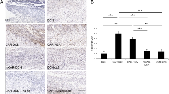
Fig. 3.
Homing of CAR–decorin in wound tissue. Mice with full thickness skin wounds received an i.v. injection of His-tagged fusion proteins on day 5 after wounding. After 4 h, the location of decorin was determined with an anti–His-tag antibody (brown). (A) The wounds of mice injected with nontargeted decorin (DCN) or mCAR–DCN were weakly positive, whereas strong staining was observed in CAR–DCN wounds. The accumulation of CAR–HSA in the wounds confirmed the ability of the CAR peptide to enhance targeting to the wounds. No staining was observed in normal skeletal muscle underlying the skin wounds of mice treated with any of the decorins (shown for CAR–DCN; CAR–DCN/muscle). No staining was seen in wound tissue when class-matched mouse IgG was substituted for the anti–6-histidine tag antibody (no ab). (Scale bar, 200 μm.) (B) Quantitative analysis of homing in skin wounds. The statistical significance was examined with ANOVA; *P < 0.05, **P ≤ 0.01, ***P < 0.001, n = 16 per group. The results are expressed as mean ± SD.

