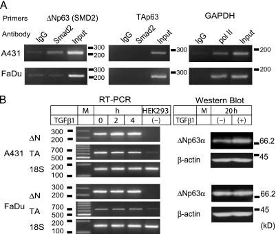
Figure 4.
Endogenous ΔNp63 activation by TGF-β1 in A431. (A) ChIP. A431 and FaDu nuclear extracts were examined for association of Smad2 with the SMD2 site. Antibodies used and amplified chromosomal segments, SMD2 (220 bp), TA (282 bp), and GAPDH (166 bp) are indicated. (B) Induction of endogenous ΔNp63 by TGF-β1. Cells incubated with TGF-β1 (10 ng/ml) for indicated periods (h) were analyzed for TAp63 and ΔNp63 mRNA by RT-PCR. Total RNA from HEK293 was tested as a negative control of ΔNp63 expression. Ribosomal RNA (18S) was amplified as a quantitative control. A 100-bp DNA ladder was loaded in lane M. Cells incubated with (+) or without (-) TGF-β1 (for 20 hours) were also analyzed by Western blot analysis for ΔNp63α. The blots were reprobed with an anti-β-actin antibody. Sizes (kDa) and positions of the standard proteins are indicated.

