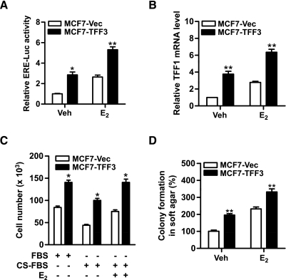
Figure 5.
Forced expression of TFF3 increases ER transcriptional activity in mammary carcinoma cells. (A) ERE-luciferase assay: MCF7-Vec and MCF7-TFF3 cells were transiently cotransfected with of pGL2-ERE-luciferase plasmid and pcDNA3 β-galactosidase plasmids in six-well plates in phenol red-free RPMI medium supplemented with 1% CS-FBS. Twenty-four hours later, the cells were treated with either DMSO (vehicle) or 10 nM E2 for another 24 hours. The cell lysates were collected and assessed for luciferase activity. (B) MCF7-Vec and MCF7-TFF3 cells were treated with either DMSO (vehicle) or 10 nM E2 in phenol red-free RPMI medium supplemented with 1% CS-FBS for 24 hours before RNA isolation. TFF1 mRNA levels were measured by qPCR. (C) Total cell number: MCF7-Vec and MCF7-TFF3 cells were cultured in 10% FBS, 10% CS-FBS, or 10% CS-FBS containing 10 nM E2 for 48 hours. (D) Colony formation in soft agar: MCF7-Vec and MCF7-TFF3 cells were seeded in 0.35% agar in a 96-well plate and treated with vehicle (Veh) or 10 nM E2 in 10% CS-FBS medium for 10 days. *P < .05, **P < .01 compared with vector control group for the same treatment.

