
Figure 1.
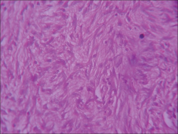
H and E stained, ×40 section shows cytologically bland spindle cells in fascicles
Official websites use .gov
A
.gov website belongs to an official
government organization in the United States.
Secure .gov websites use HTTPS
A lock (
) or https:// means you've safely
connected to the .gov website. Share sensitive
information only on official, secure websites.
H and E stained, ×40 section shows cytologically bland spindle cells in fascicles