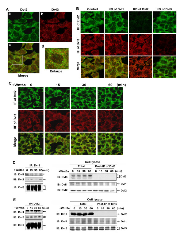
Figure 3.
Dvl2 and Dvl3 form punctate complexes. A, Fz2 expressing cells were cultured on a cover-glass in a MatTak culture dish. Cells were fixed and Dvl2 and Dvl3 were doubly stained by using antibodies against Dvl2 or Dvl3. B, cells were treated with siRNA targeting individual Dvl isoforms for 2 days and cultured on a cover-glass in a MatTak culture dish. Cells were fixed and Dvl2 and Dvl3 were doubly stained by using antibodies against Dvl2 or Dvl3. C, cells were treated with Wnt5a for indicated time periods. Cells were fixed and cellular Dvl2 and Dvl3 were doubly stained by using specific antibodies against Dvl2 or Dvl3, respectively. D and E, cells were treated with Wnt5a for indicated time periods. Immunoprecipitation-based "pull-downs" from whole-cell lysates were performed by using specific antibodies that either recognizes only Dvl3 (panel D), or recognizes only Dvl2 (panel E). The Dvl-based complexes isolated in the pull-downs ("IP:Dvl3" and "IP:Dvl2"; left panels in D and E, respectively), whole-cell lysates ("Total"; right panel), as well as supernatant fraction following the immune precipitation ("post-IP of Dvl3" and "post-IP of Dvl2"; right panels in D and E, respectively) were subjected to SDS-PAGE and the resolved proteins transferred to nitrocellulose blots. The blots were stained with antibodies specific for each Dvl isoform. A set of representative images of immunoblotting are shown.

