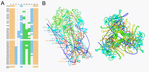
Figure 3.
Glycosylation of H1N1 viruses, 1918-2009. A) HA glycosylation status of selected, relevant H1N1 viruses from 1918 to 2009 is shown. Beige coloration shows potential glycosylation sites in the fusion domain at positions 21, 33, 289, and 154 (HA2). Blue coloration represents glycosylation status at positions 63, 81, 94, and 271 in the vestigial esterase domain. Green coloration indicates glycosylation status at positions 129 (or 131), 156, and 163 on the globular head near the receptor binding site. The site labeled 129 had the attachment asparagine at position 131 through 1984, but at position 129 from 1986 to the present; these overlapping sites are mutually exclusive so are represented together. B) Structural representations of the H1N1 HA trimer based on the crystal structure of the 1918 pandemic strain [27]. Sites for potential glycosylation have been superimposed on the structure in light blue and labeled to correspond to the chart in Figure 3A.

