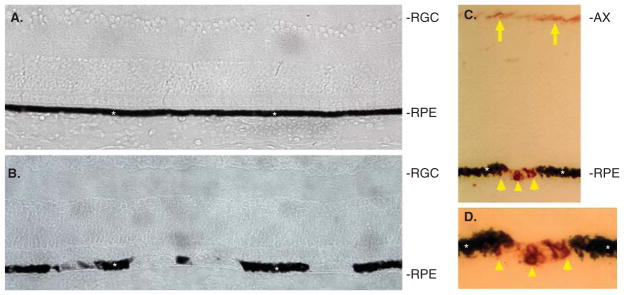
Figure 5. Sox2 induced the loss of pigmentation and the presence of RA4+ cells in the RPE layer of chick eyes.
A: Cross-section of a control E15 retina infected with RCAS-GFP. B: Cross-section of an experimental E15 retina infected with RCAS-Sox2. C: RA4 immunostaining of an experimental retina. D: Higher magnification of C. Asterisks mark the RPE. Arrows point to RA4+ axons of retinal ganglion cells. Arrowheads point to RA4+ cells in the RPE layer.
AX: Axons of retinal ganglion cells; RGC: Retinal ganglion cells; RPE: Retinal pigment epithelium.

