
Figure 1.
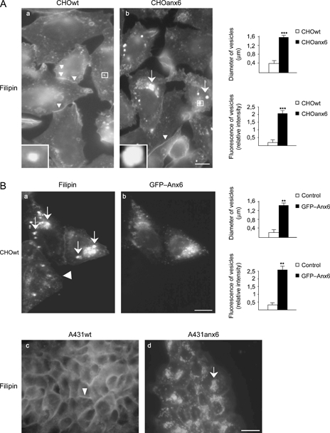
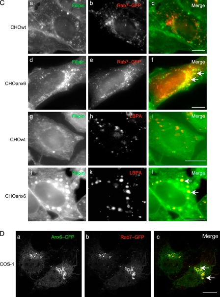
Sequestration of cholesterol in the late endosomal compartment of AnxA6-expressing cells.A) The CHOwt and CHOanx6 cells grown on coverslips were fixed and stained with filipin (5 μg/mL) as indicated. Arrows and arrowheads point at the accumulation of cholesterol in perinuclear compartments and at the PM, respectively. B) The CHOwt cells transiently transfected with GFP–Anx6 (a and b), AnxA6-deficient A431 cells (A431wt) and A431 cells stably expressing AnxA6 (A431anx6) were fixed and stained with filipin (c and d). The arrows point at the accumulation of cholesterol in perinuclear vesicles of GFP–Anx6 expressing CHO and A431anx6 cells, compared with the non-transfected control (arrowheads). For the quantification in A (a and b) and B (a and b), see text for further details. The mean values ± SD of the diameter and fluorescence intensity of perinuclear filipin-positive structures from three independent experiments (150 cells in total) per cell line is given; ** and ***, p < 0.01 and p < 0.001 for Student’s t-test, respectively. Inserts (in A) show representative filipin-positive structures. C) The CHOwt and CHOanx6 cells were transfected ± Rab7–GFP (a–c and d–f), fixed and immunolabeled with anti-LBPA (g–i and j–l) and stained with filipin (a, d, g and j) as indicated. The merged images (c, f, i and l) show a colocalization of cholesterol and LE in CHOanx6 cells (arrows in f and l). D) The COS-1 cells were cotransfected with Anx6–CFP (a) and Rab7–GFP (b), fixed and analyzed by confocal microscopy. Arrows in the merged image (panel c) point at colocalization of Anx6–CFP and Rab7–GFP. Bar is 50 μm in B (a and b) and 10 μm in all other panels. Figure 1 continued on next page.