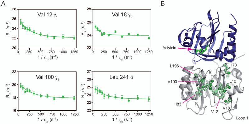
Figure 3. NMR characterization of μs-ms motions in acivicin bound ILV 13CH3 methyl labeled HisF.
A) Four representative 13C1H MQ dispersion curves out of the 17 with positive relaxation dispersion amplitudes. B) Structural mapping of ILV dispersion onto IGPS. Acivicin is shown bound to HisH in green sticks. Residues exhibiting dispersion are shown in light green spheres. Individual atoms with dispersion are highlighted with bright green spheres. See also Supplemental Table 2.

