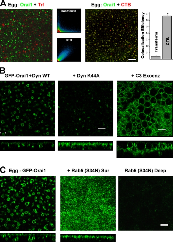
Figure 2.
Orai1 internalization endocytic pathway. (A) Oocytes expressing GFP-Orai1 were stained with 125 µg/ml Alexa Flour 633 transferrin (Trf) or 5 µg/ml Alexa Fluor 555 CTB at the GVBD stage and allowed to complete maturation. Colocalization efficiency was measured using ZEN2008 (n = 14). Error bars indicate mean ± SEM. (B) Wild-type dynamin (WT Dyn) or the K44A mutant (30 ng/cell) were injected in GFP-Orai1–expressing cells and allowed 24 h to express before inducing maturation. C3 exoenzyme (1.7 ng/cell) was injected into GFP-Orai1–expressing cells 1 h before maturation. Images are from a focal plane ∼2-µm deep from the cell surface. Orthogonal sections are also shown. (A and B) Bars, 10 µm. (C) Oocytes were injected with GFP-Orai1 alone or with 20 ng of the dominant-negative Rab5 (S34N) and then matured. Images show a deep (∼2 µm into the cell) focal plane (left) or the cell surface as indicated, with the corresponding orthogonal sections. Bar, 5 µm.

