Figure 8.
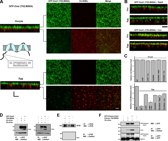
Identification of a Cav-binding site in the Orai1 N terminus. (A) The Cav consensus–binding site is illustrated in the cartoon. Cells expressing GFP-Orai1-Y52,W55A and Cherry-KDEL are shown. Planes at which the images were taken along the z stack are indicated in the orthogonal section. (B) Expression of Rab5 or Cav with Orai1-Y52,W55A does not restore its ability to internalize during meiosis. (C) Relative cell membrane Orai1 was measured as the percentage of GFP fluorescence signal on plasma membrane versus total (n = 9–38). WT, wild type. Error bars indicate mean ± SEM. (D) Orai1 was immunoprecipitated (IP) using anti-GFP antibodies from oocytes expressing 10 ng human (Ch-hCav) or Xenopus (Ch-xCav) Cav-1 with wild-type Orai1 or GFP-Orai1-Y52,W55A. Con indicates uninjected cells. Western blots (WB) were performed using anti-DsRed antibodies. The Cav band is indicated, also shown is a nonspecific band (∼33 kD) that reacts on the Western blot as a loading control. (E) Immunoprecipitation of the IP3 receptor (IP3R) as a nonspecific antigen does not pull down Cav, showing the specificity of the anti-GFP immunoprecipitation. (F) Oocytes were injected with GFP–N-term–Orai1, which contains residues 1–90 of Orai1. Cells were injected with wild-type GFP–N-term–Orai1 or a mutant with residues Y52 and W55 mutated to Ala (N-term–Y52,W55A). Immunoprecipitation was performed with an anti-GFP antibody and Western blots with either an anti-DsRed or anti-GFP as indicated. A Western blot of the different treatments is also shown (top). (D and F) Black lines indicate that intervening lanes have been spliced out. Bars, 5 µm.

