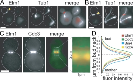
Figure 2.
Localization of Elm1. Images were collected using a wide-field microscope. Arrowheads point to Elm1-tdimer2 at the bud neck. Dashed lines indicate unbudded cells, and solid lines indicate budded cells. (A) Elm1 localizes to the bud neck. Tandem RFP/tdimer2 was fused to the carboxy terminus of Elm1 by integration at the endogenous ELM1 locus in arp1Δ mutant cells expressing GFP-Tub1. Strain: yJC6852. (B) Elm1 localizes to the neck in cells containing misoriented anaphase spindles. Strain: yJC6852. (C) Localization of Elm1 with respect to the septin network in cells expressing Elm1-tdimer2 and Cdc3-GFP. Merge image depicts Elm1 (red) and Cdc3 (green). Strain: yJC6848 with plasmid pBJ1488. (D) Quantification of normalized fluorescence intensities for Elm1, Cdc3, Bni4, and Kcc4 across the bud neck. Images were collected from cells simultaneously expressing Elm1-tdimer2 and either Cdc3-GFP, Bni4-GFP (yJC7251), or Kcc4-GFP (yJC7252). Fluorescence intensities were measured across a 2-µm region, depicted in C, extending from the mother into the bud and centered on the smallest diameter of the neck (dashed line). Intensities were measured in at least 10 large-budded cells. Values for Elm1 are compiled from the three strains, totaling 50 cells. Fluor intensity values are given in arbitrary units. Bars, 2 µm.

