Figure 2.
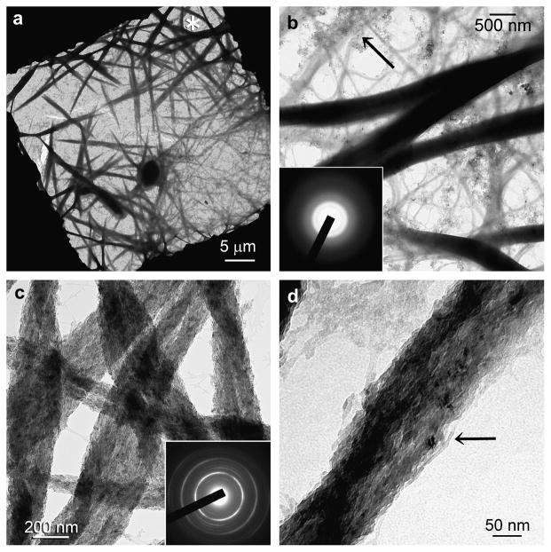
Unstained TEM showing the temporal events associated with non-hierarchical intrafibrillar mineralisation in the sequestration analogue control (no templating analogue). (a) Grid space showing swollen electron-dense collagen fibrils (asterisk) after 24 hours. (b) Swollen, electron-dense fibrils with a smooth appearance. ACP nanoparticle clusters were seen predominantly around unmineralised fibrils (arrow). ACP nanoparticles appeared to have penetrated the swollen fibrils and coalesced into a continuous amorphous mineral phase (inset). (c) After 72 h of mineralization, collagen fibrils were no longer swollen and exhibited a fibrous appearance. Although they contained intrafibrillar mineral, they lacked cross-banding patterns. Inset: Selected are electron diffraction (SAED) of individual mineralised fibrils produced ring patterns that are characteristic of apatite (note arc-shaped patterns indicating that minerals are arranged along the longitudinal axis of the collagen fibrils – see also Supplementary Fig.4). (d) High magnification of a mineralised fibril showing the continuity of the mineral strands and absence of discrete crystallites (arrow).

