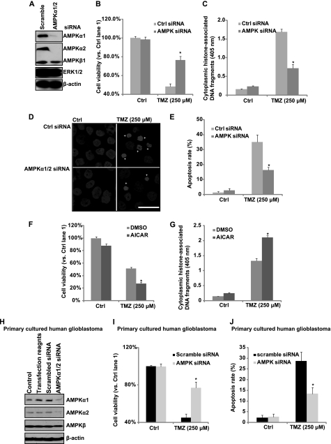
FIGURE 3.
AMPK activation is involved in TMZ-induced cell death in vitro. A, glioblastoma U87MG cells were transfected with control (scrambled) or AMPKα1/2 siRNA for 48 h. AMPKα1/2, AMPKβ, ERK1/2, and β-actin expression levels were detected by Western blotting. Successfully AMPKα knocked down cells were used for further experiments. U87MG cells transfected with control or AMPKα siRNA were treated with 250 μm TMZ. B, cell viability was detected by MTT assay after 48 h. C–E, cell apoptosis was detected by histone/DNA ELISA (C) and Hoechst 33342 staining (D and quantified in E) after 36 h. F and G, U87MG cells were pretreated with AICAR (1 mm) followed by 250 μm TMZ treatment. Cell viability was detected by MTT assay after 48 h (F). Cell apoptosis was detected by histone/DNA ELISA after 36 h (G). H, primary cultured human glioblastoma cells were either left untreated or transfected with control (scramble) or AMPKα1/2 siRNA for 48 h. The expression level of AMPKα1/2, AMPKβ, and β-actin were detected by Western blotting. Successfully AMPKα knocked down cells were used for further experiments. DMSO, dimethyl sulfoxide. I, primary cultured human glioblastoma cells transfected with control or AMPKα siRNA were treated with 250 μm TMZ. Cell viability was detected by MTT assay after 48 h. J, cell apoptosis was detected by Hoechst 33342 staining after 36 h. Experiments in this figure were repeated at least three times, and similar results were obtained. *, p < 0.05 versus TMZ-treated group in control group (ANOVA). Scale bar, 25 μm.

