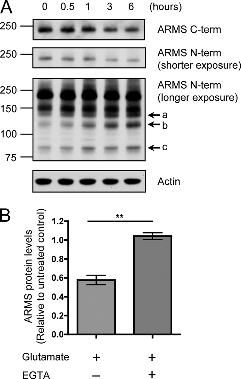
FIGURE 4.

Glutamate activation of hippocampal neurons induces ARMS/Kidins220 degradation through a calcium-mediated process. A, ARMS/Kidins220 is degraded upon glutamate treatment of hippocampal neurons. Hippocampal cultures were stimulated with 200 μm glutamate for 1 min, and cell lysates were collected at the indicated times after stimulation and immunoblotted for ARMS/Kidins220. As was the case after KCl depolarization, full-length ARMS/Kidins220 was degraded, leading to the accumulation of similar smaller sized N-terminal fragments (arrows a, b, and c). Actin is shown as a loading control. A 150-kDa nonspecific band was detected by the N-terminal antibody. B, glutamate-mediated ARMS/Kidins220 degradation requires extracellular calcium. Hippocampal cultures were stimulated with 200 μm glutamate for 1 min in the presence of EGTA, and cell lysates were collected 3 h after stimulation. ARMS/Kidins220 protein levels, as detected by the C-terminal antibody, were assessed by Western blot and quantified. ARMS/Kidins220 levels were normalized to actin levels and plotted relative to untreated control (n ≥ three independent experiments; **, p < 0.01, t test; data are represented as the means ± S.E.).
