FIGURE 4.
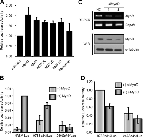
MyoD enhances SelW gene promoter activity. A, activity of the SelW promoter by MRFs and MEF2 factors. Nonmyogenic C3H10T1/2 cells were co-transfected with −973SelW/Luc and pRL-TK, together with an expression plasmid containing a MRF (MyoD, Myf5, and myogenin) or MEF2 (MEF2A, MEF2C, and MEF2D) factor. The cells were cultured in GM and harvested for Dual-Luciferase reporter analysis. Results are expressed as means ± S.D. of four independent experiments in triplicate. B, effect of exogenous MyoD on SelW gene promoter activity. SelW promoter plasmids and 4RSV-Luc plasmid were co-transfected with or without an expression plasmid containing MyoD into C3H10T1/2 cells. The cells were cultured in GM up to ∼100% confluence, maintained, and harvested after 1 day of culture in DM. Results are expressed as means ± S.D. of four independent experiments performed in triplicate. C, knockdown of MyoD expression by siRNA in C2C12 cells. C2C12 myoblasts were separately transfected with two distinct siRNAs specific for MyoD (siMyoD 1 and 2) along with a negative control (NC). The cells were maintained in GM up to ∼90% confluence, followed by cultivation in DM for 1 day. MyoD expression was monitored by RT-PCR and Western blot analysis. The Gapdh gene and α-tubulin protein were used as loading controls for each analysis. D, effect of MyoD depletion on SelW promoter activity in differentiating C2C12 cells. C2C12 cells transfected with siMyoD 2 were analyzed for SelW gene promoter activity after 1 day of culture in DM. Data are represented as means ± S.D. of four independent experiments performed in triplicate.

