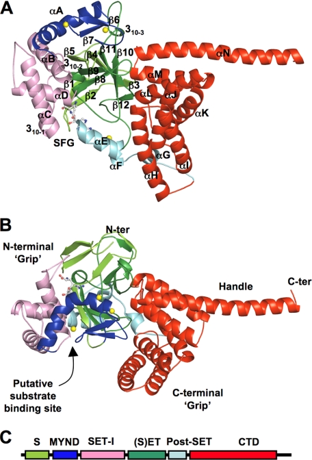
FIGURE 1.
Ribbon diagram of the overall structure of SmyD1 in complex with sinefungin. A, top, and B, side views. Secondary structures of SmyD1, α-helices, 310-helices, and β-strands are labeled and numbered according to their position in the primary sequence. The S-sequence, MYND, SET-I, core SET, post-SET, and CTD domains are depicted in light green, blue, pink, green, cyan, and red, respectively, whereas sinefungin (SFG) is represented by ball-and-stick and zinc ions by yellow spheres. Sinefungin is a structural analog of AdoMet and a potent inhibitor of AdoMet-dependent methyltransferases. Using sinefungin instead of AdoHcy or AdoMet in co-crystallization was necessary to obtain diffraction quality crystals. C, schematic diagram of SmyD1 domain structures.

