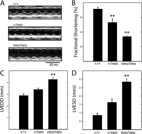
FIGURE 7.
Echocardiographic analysis of hearts from mice of indicated genotype. A, representative M-mode echocardiograms recorded in unanesthetized mice. Details of the analysis are shown in the larger images in Fig. 8. B, left ventricular fractional shortening ((LVEDD − LVESD)/LVEDD) expressed as percent. C, LVEDD. D, LVESD. Data were obtained on 18- to 22-week-old male mice. n = 9, 6, and 15 for cMLCK+/+, MLCK+/neo, and MLCKneo/neo mice, respectively. **, p < 0.01 compared with cMLCK+/+ mice.

