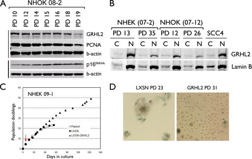
FIGURE 1.
GRHL2 expression is decreased during senescence of NHK. A, Western blotting was performed with the WCEs of primary NHOK (strain 08-2) harvested at the indicated PD levels. Intracellular levels of GRHL2, PCNA, and p16INK4A were determined. β-Actin was probed for the loading control. B, NHEK (07-2) and NHOK (07-12) cultures were fractionated into cytoplasmic (denoted as C) or nuclear (denoted as N) samples at the indicated PD levels. Western blotting was performed for GRHL2 and lamin B. Fractionated samples of SCC4 were included for comparison. C, rapidly proliferating NHEK (strain 09-1) were infected with LXSN or LXSN-GRHL2 and maintained in serial subcultures. Red arrow indicates the point of infection with LXSN-GRHL2. D, NHOK infected with LXSN or LXSN-GRHL2 were stained for senescence-associated β-galactosidase activity at the indicated PD levels, which were calculated from the point of primary culture. Original magnification, ×100.

