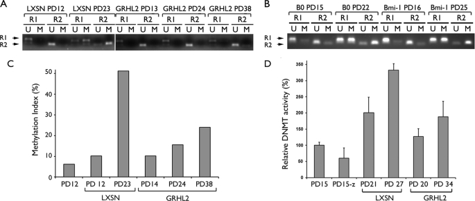
FIGURE 8.
GRHL2 inhibits DNA methylation of the hTERT promoter. A, MSP was performed with the bisulfite-modified genomic DNA isolated from the cells at the indicated PD levels. The NHEK/LXSN cells (09-1) were rapidly proliferating at PD 12 and senesced at PD 25, and the NHEK/GRHL2 cells proliferated exponentially and entered the senescing phase near PD 38 (Fig. 1). R1 and R2 indicate the two different regions of the hTERT promoter amplified by MSP (see “Experimental Procedures”). U and M indicate amplification with the primer sets recognizing the hypo- and hypermethylated DNA, respectively. B, MSP was also performed with the NHK/B0 cells harvested at PD 15 (replicating) and PD 22 (senescing) and with actively replicating NHK/Bmi-1 cells harvested at PDs 16 and 25. C, MSP was performed with quantitative PCR for the R2 region, and the methylation index was calculated by taking the % ratio of the M amplification signal to the combined M and U signals. D, DNMT1 enzyme activity was performed with the nuclear extracts of NHEK (PD 15) and the cells infected with LXSN (PDs 21 and 27) and LXSN-GRHL2 (PDs 20 and 34). The parent NHEK (PD 15) treated with 5-aza-CdR (1 μm) for 5 days was included as a control (denoted as PD15-z).

