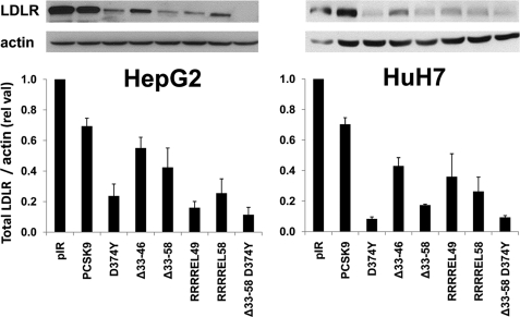
FIGURE 3.
Effect of extracellular PCSK9 and its GOF mutants on total LDLR levels in HepG2 cells. Western blot analysis of LDLR in HepG2 (left panel) and HuH7 (right panel) cells transfected with PCSK9 and its gain of function mutants D374Y, Δ33–46, Δ33–58, and D374Y-Δ33–58. The estimated % decrease in total LDLR was normalized to β-actin levels. These data are representative of three independent experiments.

