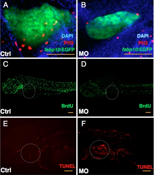
FIGURE 5.
Knockdown of GrnA impairs liver cell proliferation and enhances apoptosis. Control (Ctrl) and grnA MO-injected embryos were examined using the anti-PH3 antibody, BrdU incorporation, and a TUNEL assay at 4 dpf. DAPI was used to stain the cell nuclei. Fewer PH3-positive cells were detected in MO-injected Tg (fabp10:EGFP) embryos compared with control MO-treated embryos at 4 dpf (A and B). BrdU incorporation was suppressed in the liver in grnA morphants compared with controls (C and D). TUNEL staining of control MO-injected embryos (E) and knockdown embryos (F). A dotted line circles the liver. Scale bars, 100 μm.

