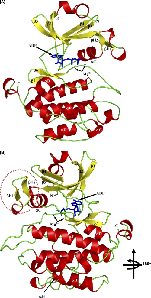
FIGURE 2.
Crystal structure of the kinase domain of PASK. A, secondary structure elements of PASK kinase domain in the presence of ADP are shown. The α-helices are depicted in red, β-strands in yellow, loops in green, and ADP and Mg2+ ions in blue. The strands and helices are named according to the conventional nomenclature for PKA. The N and C termini of the kinase structure are indicated as N and C, respectively. B, PASK three-dimensional structure is rotated 180° along the vertical axis to show the positions of βH1 and βH2 helices (marked). See “Results” for details.

