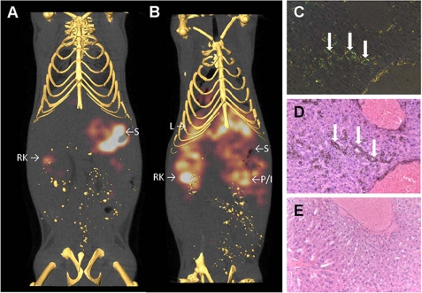
FIGURE 8.
Imaging, and specific co-localization of NS4F5 with AA amyloid deposits in vivo. The distribution of 125I-labeled NS4F5 was visualized in healthy (A) and amyloid-laden (B) mice by microSPECT imaging. The antibody accumulated in amyloid deposits in the liver (L) as well as the pancreas (or intestine, P/I) and kidney (RK for right kidney). In amyloid-free mice, liberated radioiodide was observed in the stomach (S) with some activity also associated with renal clearance of the antibody (RK). Specific uptake of 125I-labeled NS4F5 by hepatic amyloid deposits was confirmed microscopically, where Congo-philic amyloid (C) was co-stained by black grains in the autoradiograph (D). There was no accumulation of 125I-labeled NS4F5 in a healthy liver (E) (n = 4).

