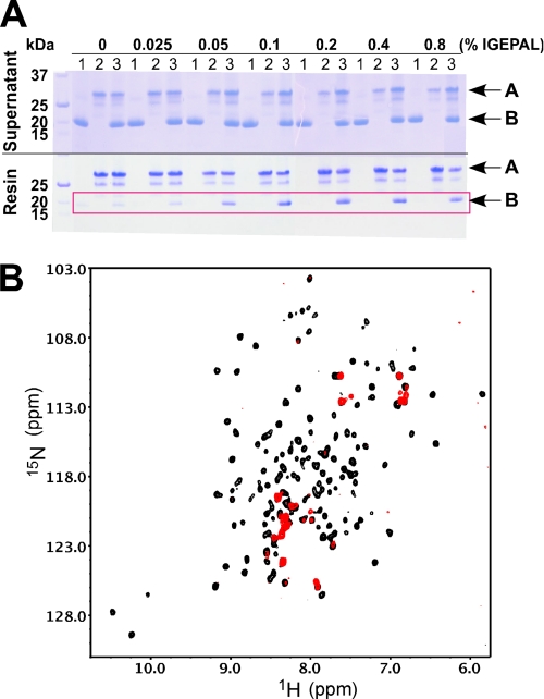
FIGURE 5.
Interaction between BAK and MCL-1 in the presence of IGEPAL. A, Coomassie-stained SDS-gels showing the supernatant (top) and proteins retained by the Ni2+-NTA resin (bottom) under the conditions indicated. Conditions 1–3, respectively, mean that cMCL-1, FLAG-BAK-HMK-His6, or their combination was loaded. Arrows labeled A show the position of FLAG-BAK-HMK-ΔTM-His6, and arrows labeled B show the position of cMCL-1. The red frame highlights the cMCL-1 retained by FLAG-BAK-HMK-ΔTM-His6 in the presence of IGEPAL. B, 1H-15N HSQC spectra of 15N-labeled cMCL-1 in 0.1% IGEPAL without (black) and with (red) unlabeled cBAK.

