
Figure 1.
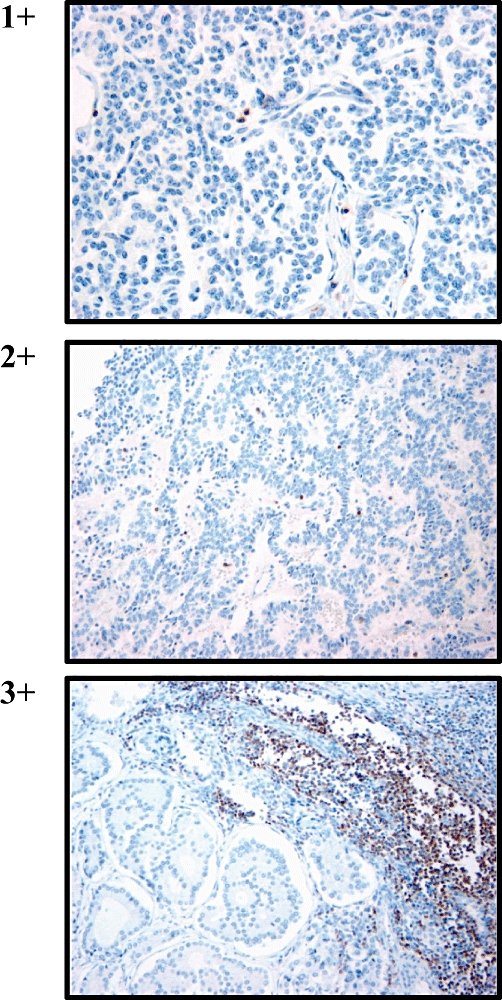
Neuroendocrine tumours were stained with anti-CD3 to identify infiltrating T cells. Grading levels were assigned according to the following criteria per 10 highpower fields: grade 0 = < 10 cells; grade 1 = < 1% or 10–20 cells; grade 2 = 1–5% or 21–50 cells, and grade 3 = > 5% or >50 cells. (Haematoxylin and eosin stain; original magnification 400×)