
Figure 2.
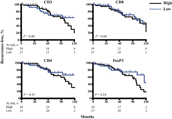
Neuroendocrine tumours were stained with anti-CD3 (all T cells), anti-CD8 (cytotoxic T cells), anti-CD4 (helper T cells), and anti-FoxP3 (regulatory T cells) to quantify the numbers of various T cell subsets within the tumours. Kaplan–Meier analyses were performed to determine differences in recurrence-free survival among patients with high and low levels of T cell infiltration