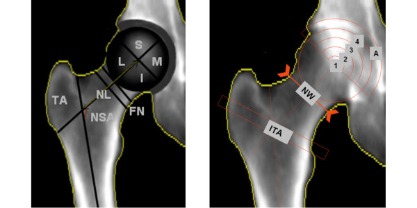
Figure 1.
DXA images that show the parameters that are determined in the software for the DXA scan analysis. a) Trochanteric area (TA), Neck shaft angle (NSA), femoral neck length (NL): line from the center of the femoral head to the intersection point of the femoral shaft and femoral neck (FN). The femoral head was divided in four quarters: Superior (S), Medial (M), Inferior (I), and lateral (L). b) Arcs dividing the upper part of the femoral head in four sub regions ranging from the center of the subchondral region and acetabular area (A), neck width (NW) measured on the narrowest neck region and intertrochanteric area (ITA). For all areas the BMD, BMC and area size were determined.

