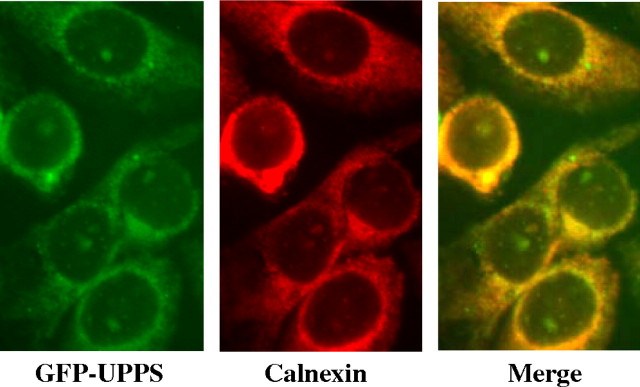
Fig. 5.
ER localization of GFP-UPPS expressed in CHO cells by immunofluorescence. CHO cells expressing E. coli UPPS with an N-terminal GFP tag (left, green) were viewed, with and without treatment with rabbit anticalnexin antibody, followed by Texas Red conjugated antirabbit antibody (center, red), with a Nikon Eclipse E600 microscope, as described in “Materials and methods”. The overlap image (right, merge) indicates colocalization of GFP-UPPS with calnexin.

