Figure 1.
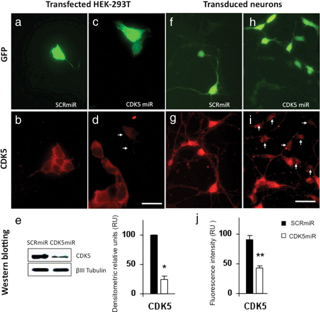
Knockdown of CDK5 in HEK-293T cell line and neuronal primary cultures by CDK5miR constructs. a, GFP expression of HEK-293T cells transfected with pCMV-GIN-ZEO-shRNAmirSCR.GFP (SCRmiR). b, CDK5 immunofluorescence in HEK-293T cells transfected with SCRmiR. c, GFP expression of HEK-293T cells transfected pCMV-GIN-ZEO-shRNAmirCDK5.GFP (CDK5miR). d, CDK5 immunofluorescence in HEK-293T cells transfected with CDK5miR; the arrows point to GFP+ cells in c. Decrease of the CDK5 immunoreactivity is observed in transfected cells. Green, GFP fluorescence; red, Alexa 594. Magnification, 60×. Scale bar, 20 μm. n = 6. e, CDK5 Western blotting in HEK-293T transfected cells with SCRmiR and CDK5miR. A representative plot is shown. βIII-Tubulin was used as loading control. Data are presented as mean ± SEM. n = 6. *p < 0.05. f, GFP expression of neuronal primary cultures transduced with LV- SCRmiR. g, Immunofluorescence of CDK5 in transduced neurons with LV-SCRmiR. h, GFP expression of neuronal primary cultures transduced with LV-CDK5miR. i, CDK5 immunofluorescence in neurons transduced with LV-CDK5miR. The arrows point to GFP+ transduced neurons seen in h. Decrease of CDK5 immunoreactivity in transduced neurons with LV-CDK5miR is observed. Green, GFP fluorescence; red, Alexa 594. Magnification, 60×. Scale bar, 20 μm. n = 6. **p < 0.001. j, Quantification of the fluorescence intensity of CDK5 immunoreactivity of neurons transduced with LV-SCRCDK5miR and LV-SCRmiR, using the software Image Scope-Pro (Media Cybernetics). RU, Relative units. n = 6. **p < 0.001.

