Figure 5.

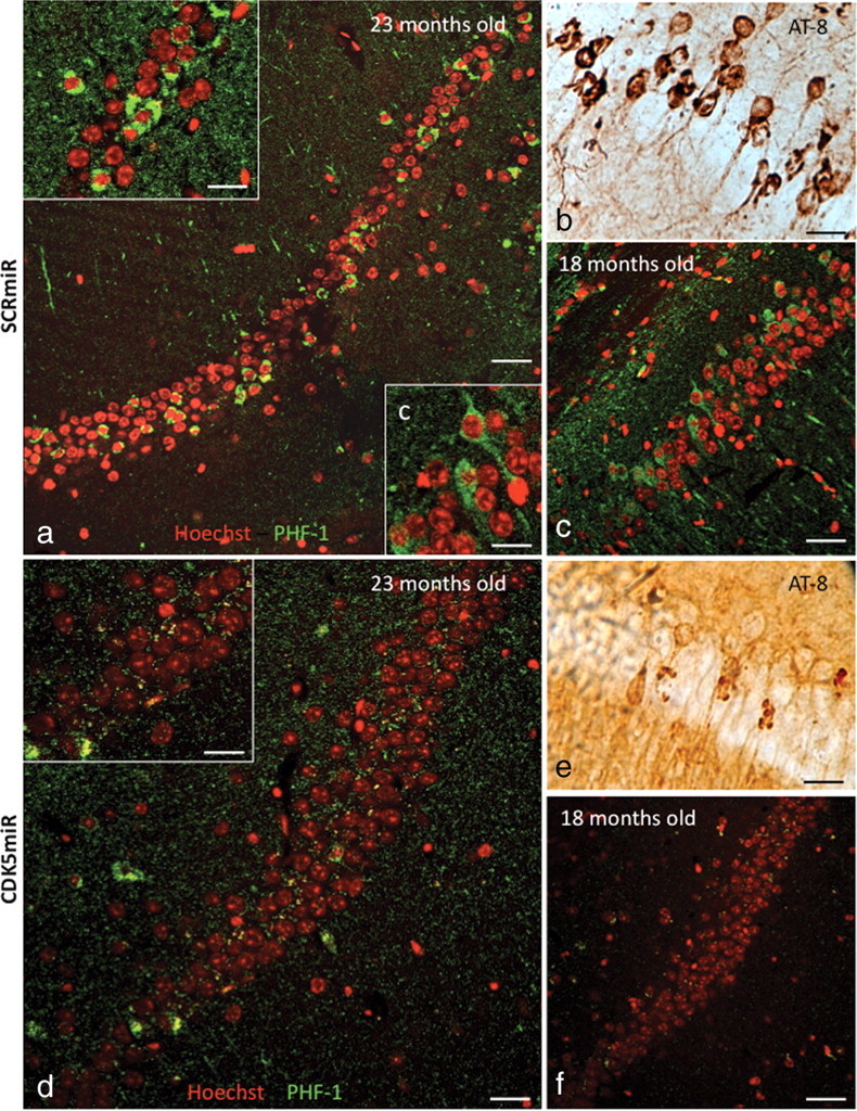
CDK5miR decreases phosphorylated tau and neurofibrillary tangles in 3×Tg-AD mice. a, b, Phospho-tau (PHF-1) immunofluorescence showing NFT-positive cells (a) and phospho-tau (AT-8)-immunoreactive cells (b) present in the CA1 area of hippocampus of 23-month-old 3×Tg-AD mice 3 weeks after injection with AAV-SCRmiR. c, PHF-1 immunofluorescence showing positive cells in CA1 in hippocampus of 18-month-old 3×Tg-AD mice, 3 weeks after hippocampal injection with AAV-SCRmR. d–f, PHF-1 immunofluorescence (d) and AT-8-immunoreactive cells in the CA1 area of 23-month-old (e) and 18-month-old (f) 3×Tg-AD mice 3 weeks after injection with CDK5miR. n = 6. Red pseudocolor: Nucleus staining with Hoechst; green pseudocolor: PHF-1 IF, Alexa 594. Magnification, 60×. Scale bar, 50 μm. Magnification: insets a, c, d, 100×. Scale bar, 20 μm.
