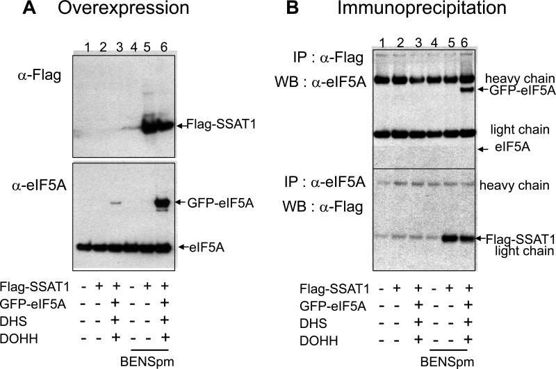
Figure 5. Physical interaction between eIF5A and SSAT1 in cells.
HeLa cells were cotransfected with GFP-eIF5A, DHS, DOHH and FLAG-SSAAT1 vectors as indicated and treated with or without 10 μM BENSpm, which was added at the time of medium change after transfection. (A) The overexpression of Flag-SSAT1 and GFP-eIF5A was detected by western blotting of cell lysates. (B) The cell lysates were immuno-precipitated with either with anti-FLAG antibody (top panel) or with eIF5A antibody (bottom panel) and immuno-precipitates were analyzed by western blotting with antibodies indicated. The experiments were repeated once with consistent results.

