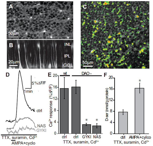
Figure 4.

AMPA+cyclothiazide D-serine release is not blocked by inhibition of neural activity. (A) Fluo-4 AM loaded retina showing labeling of Astrocytes (black arrow) and Müller cell endfeet surrounding unlabeled ganglion cell bodies (white arrow). (B) Cross-section of retina in A reconstructed from a Z-series showing Müller cell labeling of endfeet near the ganglion cell layer (GCL), with stalks spanning the inner plexiform layer (IPL), and cell bodies in the inner nuclear layer (INL). (C) Ca2+ response of Müller cell endfeet to AMPA+cylco. Retinas were bulk loaded with Fluo-4 AM then treated with 1μM TTX, 100 μM suramin, and 200 μM Cd2+ 2 minutes prior to a 30 second bath application of AMPA+cylco. Image shows the change in Ca2+ in response to AMPA+cylco as a thresholded maximal projection (pseudocolor) overlaid on the average baseline image. Ca2+ elevations excluded ganglion cell bodies. (D) Change in Ca2+ over time from the region in C. (E) No significant difference was observed in the Ca2+ increase between wt (n=4) and DAO- (n=3) retinas following AMPA+cylco application. GYKI (n=5) and NAS (n=6) significantly reduced the AMPA+cylco induced Ca2+ response in DAO- retinas (*, p<0.05, compared to ctrl). (F) DAO- retinas exposed to AMPA+cyclo still released D-serine in the combined presence of TTX, suramin, and Cd2+. (*, statistically different from control, p<0.05, n=4-5).
