Fig. 3.
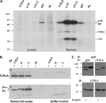
FCRLA binds intracellular IgM. (A) Jurkat T cells and Ramos B cells were metabolically labeled overnight, lysed with NP-40, cleared with protein A-sepharose beads (PA) and then immunoprecipitated with antibodies to FCRLA (M116), μHC, κ and λ LC and BiP. The samples were analyzed by SDS–PAGE under reducing conditions. The migration of molecular weight markers is indicated on the left side of the gel. (B and C) FCRLA preferentially binds to μs in Ramos B cells. (B) Ramos NP-40 cell lysates (left) or buffer only controls (right) were precleared with protein A-sepharose beads (PA) and then immunoprecipitated with the monoclonal FCRLA and polyclonal anti-Ig antibodies indicated along the top of the figure. The blots were probed with polyclonal FCRLA (a) or μHC (b) antibodies. The right side of this figure contains the precipitating antibodies but no cell lysate, a necessary negative control as the second antibodies used to develop the blots cross react with the immunoprecipitating antibodies. (C) Ramos cell lysates were immunoprecipitated with polyclonal anti-μ (left panels) and monoclonal FCRLA antibodies (right panels). Immunoprecipitates were treated with EndoH (+) or were mock treated (−). The blots were probed (IB) with anti-μ (upper panel) and then stripped and probed with anti-FCRLA (lower panel).

