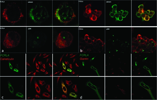
Fig. 7.
Intracellular localization of FCRLA as determined by confocal microscopy. The localization of FCRLA in BJAB (a) and transfected 293T cells (b) was determined by staining with anti-FCRLA antibody (red) and anti-calnexin or anti-p58k antibodies (green). The localization of FCRLA in transfected HeLa cells was determined by staining with anti-FCRLA Alexa 488-conjugated M101 FCRLA mAb (green) and anti-calreticulin (c) or anti-giantin (d) antibodies (red). Images of sections of the stained cells captured by a laser scanning confocal microscope are shown. Yellow color shows co-localization of FCRLA with the ER markers.

