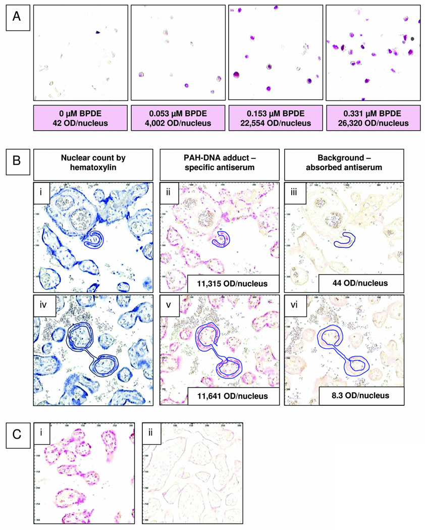
Fig. 1.
(A) Cultured human cervical keratinocytes exposed to 0, 0.053, 0.153, or 0.331-µM BPDE for 1 h were fixed, paraffin-embedded, and stained with rabbit anti-BPDE-DNA and Fast Red. Values for pink color intensity, optical density (OD)/nucleus, determined by ACIS are shown below each image. (B) Sequential sections from 2 human placenta samples were stained with either hematoxylin (i and iv), anti-BPDE-DNA antiserum (1:20,000, ii and v), or immunogen-absorbed anti-BPDE-DNA (1:20,000, iii and vi). Nuclei located in the regions designated for adduct analysis (outlined in blue with the ACIS regional selection tool) were counted on hematoxylin-stained slides. (C) Human placenta samples that were either fixed when fresh (Group 1, i), or frozen for 5 years before fixation (Group 2, ii) were stained simultaneously with anti-BPDE-DNA (1:10,000); PAH-DNA adducts were virtually undetectable in the frozen samples.

