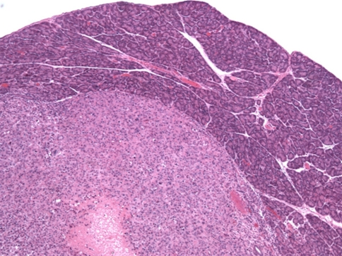
Fig. 6.
Histologic image of a resected green fluorescent protein (GFP)-expressing, orthotopic tumor implant. Hematoxylin and eosin (H&E)-stained sections demonstrated the expansile tumor formed by orthotopically implanted GFP-expressing human pancreatic carcinoma cells (MiaPaCa-2) within the body of the mouse pancreas. The poorly differentiated tumor, surrounded by a variable thickness of normal pancreatic parenchyma, showed scattered foci of zonal necrosis, as seen in the lower edge of the image

