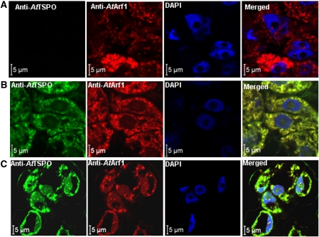
Fig. 1.
AtTSPO colocalizes with AtArf1 in plant cells. (A) Wild-type cells were probed with anti-AtArf1 and Texas red-coupled anti-rabbit secondary antibody, then with anti-AtTSPO-coupled to FITC; the cells were mounted in antifade medium containing DAPI and imaged sequentially with a confocal microscope (Zeiss LSM 710) using an oil-immersion ×63 lens. No FITC signal could be detected in wild-type cells. (B) A transgenic cell line overexpressing 6×His-ATTSPO was treated as in (A), a FITC-derived signal could be seen (panel AtTSPO) and the merged image (panel merged) showed a perfect colocalization with the Texas red-derived signal. (C) Wild-type cells were pre-incubated in ABA (50 μM) before probing as in (A); the FITC-derived signal could be seen and perfectly colocalized with the Texas red-derived signal as in (B).

