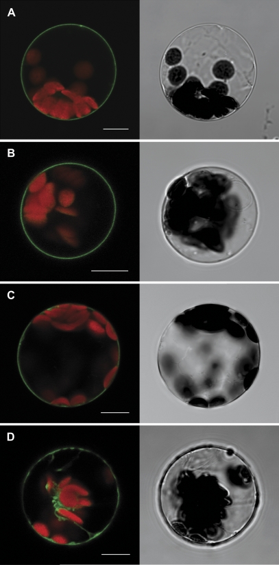
Fig. 2.
Unique localization of SAUL1 and its homologues at the plasma membrane. Transformation of Arabidopsis protoplasts followed by analyses of fluorescence signals by confocal laser scanning microscopy indicated the localization of SAUL1–GFP (A), GFP–PUB43 (B), and GFP–PUB42 (C) fusion proteins at the plasma membrane, and of GFP–PUB5 fusion proteins in the cytoplasm (D). Autofluorescence of chlorophyll is shown in red. Transmitted light pictures of the transformed protoplasts are shown next to the respective fluorescence picture. Scale bars represent 10 μm.

