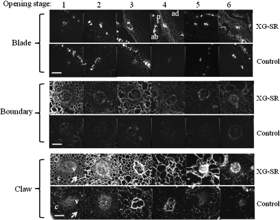
Fig. 6.
In situ detection of XET activity in carnation petal tissues by detecting fluorescence from wall-bound xyloglucan conjugated with XLLG-SR by the action of XET. Cryostat sections of 100 μm thick prepared by the thin film reinforcement method were used for observation with a fluorescence microscope. The control reaction mixture contained GGGG-SR instead of XLLG-SR. Arrows in the plate of the claw at opening stage 1 show the difference in fluorescence between the XLLG-SR-treated and the GGGG-SR-treated control tissues. Flower opening stages are similar to those described in the legend to Fig. 4. ab, abaxial epidermis; ad, adaxial epidermis; c, cell wall of parenchyma cells; p, parenchyma; v, vascular bundle. Scale bars=100 μm.

